An international team of researchers will for the first time be able to demonstrate clinical-quality Proton CT to improve Proton Therapy in the treatment of cancer – moving a step closer to this improved treatment method being used to help those suffering with certain forms of cancer, particularly for children and young people.
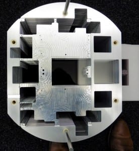
Led by Distinguished Professor of Image Engineering Nigel Allinson MBE, from the University of Lincoln, UK, the pioneering PRaVDA (Proton Radiotherapy Verification and Dosimetry Applications) project is developing one of the most complex medical instruments ever imagined to improve the delivery of proton beam therapy.
The team has time on the South African National Cyclotron (a type of particle accelerator), near Cape Town, and hopefully later this year will have a world-first in showing clinical-quality Proton CT.
Professor Allinson said: “The uncertainties in where the protons lose their energy and do damage (tumour or healthy tissue) will only be eliminated by using the same type of radiation, Protons, to image and to treat. By delivering clinical quality Proton CT images, this project will greatly improve the treatment of cancer using proton therapy. Such therapy is particularly useful in the treatment of young people, those with brain tumours, and eventually it may help such stubborn cancers as lung cancer.
“It has been an extraordinary engineering feat – certainly the most complex piece of engineering undertaken at the University of Lincoln. It has involved the active involvement of six universities, four NHS Trusts, National Research Laboratories in South Africa, two specialist sub-contractors and numerous UK and European suppliers.
“The system including detectors of the type used in the Large Hadron Collider and CMOS imagers like those to be found in your smartphone but 500 times larger and working 20 times faster. There is enough processed silicon wafers to make more than 25,000 iPhone cameras. The data output of the system is equivalent to over 300 HDTV channels.”
The innovation will assist radiotherapists by helping them to achieve accurate proton CT images. Nearly half of all cancer patients receive radiotherapy as part of their curative treatment, and most radiotherapy is delivered using high-energy external beams of x-rays. Proton beam therapy, however, uses a different type of beam to conventional radiotherapy. It uses a high-energy beam of protons. Like x-rays, protons can penetrate tissue to reach deep tumours. However, compared to x-rays, protons cause less damage to healthy tissue in front of the tumour, and no damage at all to healthy tissue lying behind, which greatly reduces the side effects of radiation therapy.
There are uncertainties in exactly where the protons will lose most of their energy and hence kill the tumour while not adversely affect healthy tissue. For a tumour 20cm deep inside a patient, this uncertainty is about 1.5 cm. Using Proton CT will eliminate all of these errors.
In November 2014 the consortium received a prestigious Institution of Engineering and Technology (IET) Innovation Award, named as the winner in the Model-Based Engineering category.
The project is funded by The Wellcome Trust.


