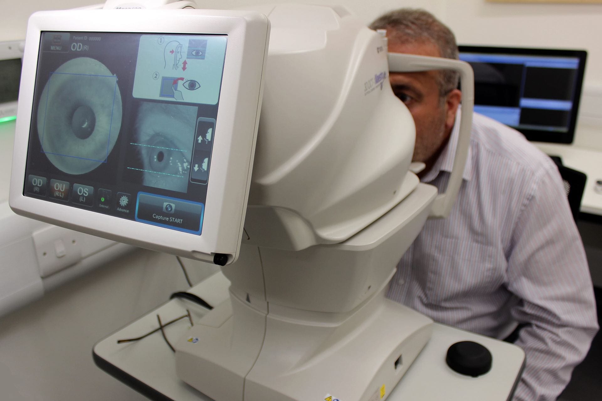
Principal Investigator, Dr Bashir Al-Diri and colleagues at the School of Computer Science are conducting a research project to collect 2D fundus images and 3D Optical Coherence Tomography (OCT) images using a fully automated user-friendly retinal-imaging camera (3D OCT-1 Maestro).
This study, titled ‘Automated Retinal Imaging Lab (ARIAL), will look towards finding and analysing new signs in the retinal vascular system photographed at the back of the eye, which might be changed due to disease. These signs can then be monitored and measured over time to detect signify disease progression.
OCT images are the most common techniques used for detecting eye diseases affecting the macula; OCT images are using in routine clinical practice and for diagnosis and monitoring diseases such as diabetes and high blood pressure as well as other systemic diseases.
All images will be reviewed and stratified by a Consultant Ophthalmologist Surgeon. Any abnormality will be reported directly to you and your registered GP with clear advice on further action if needed.
For this study, we welcome everyone with or without any known eye disease or diagnosed with any chronic systemic diseases. OCT images and lifestyle data will be captured and collected every 6 months for the duration that you are available; each visit will take no longer than 30 minutes.
There has been no such dataset available for the research community in the past, so this project will be of great scientific interest.
Further information can be found here. To participate, please contact Dr Bashir Al-Diri:
T. 01522 837111 / E. baldiri@lincoln.ac.uk


